
17/02/2026
Share:
17/02/2026
Share:
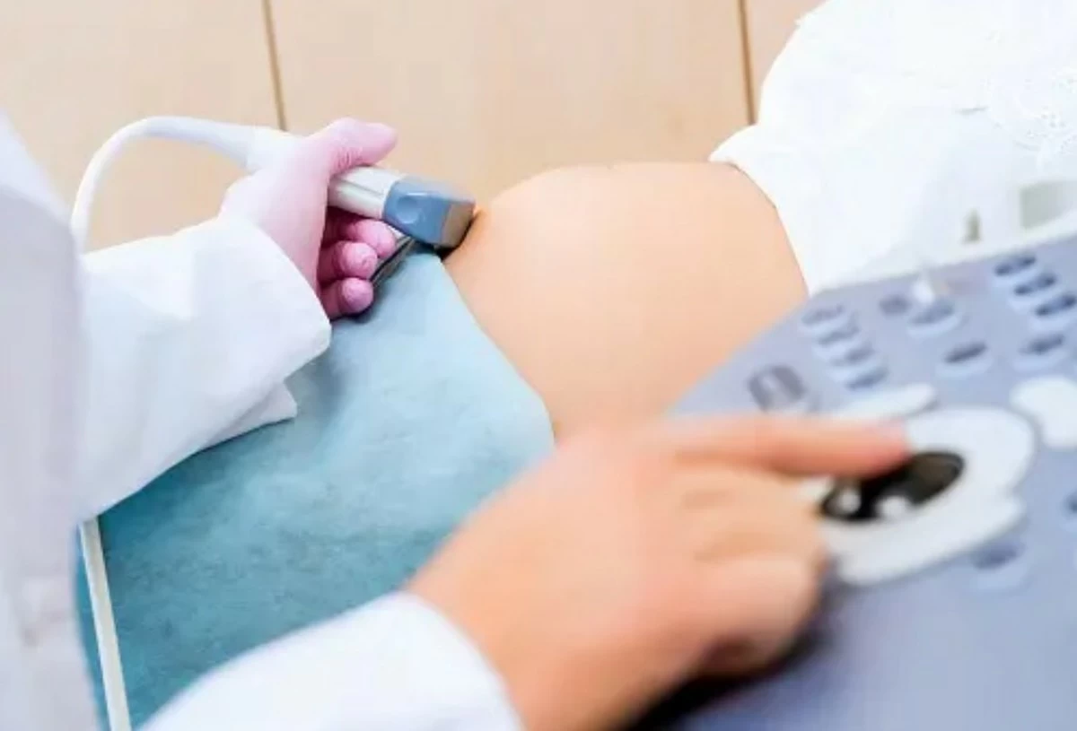
KUWAIT CITY, Feb 17: Dr. Tariq Al-Arabi, a specialist in obstetrics and gynecology, emphasized the importance of combining detailed ultrasound scans with blood tests in the early stages of pregnancy. In a press statement, Al-Arabi explained that this combination plays a vital role in ensuring the accuracy of results and medical reassurance regarding the well-being of the fetus. He said the first accurate ultrasound scan typically begins between the 11th and 14th weeks of pregnancy, and it is a crucial step in the early assessment of fetal health.
He added that the ultrasound scan measures the translucency of the fetal neck and examines the nasal bone, both of which are important indicators for the early detection of certain genetic syndromes. He added that the second stage of the detailed examination takes place between the 18th and 22nd weeks of pregnancy, known as a detailed ultrasound.
During this scan, the fetus’ organs are fully assessed, including the brain, heart, digestive system, urinary system, spine and limbs, in addition to the placenta and the amount of amniotic fluid. He pointed out that in some cases, 4D ultrasound is added and 5D ultrasound technology is also available, which is often used in the sixth and seventh months of pregnancy. He added that 5D ultrasound provides a high-resolution image combining advanced visual clarity with medical reassurance on the condition of the fetus.
By Marwa Al-Bahrawi Al-Seyassah/Arab Times Staff